
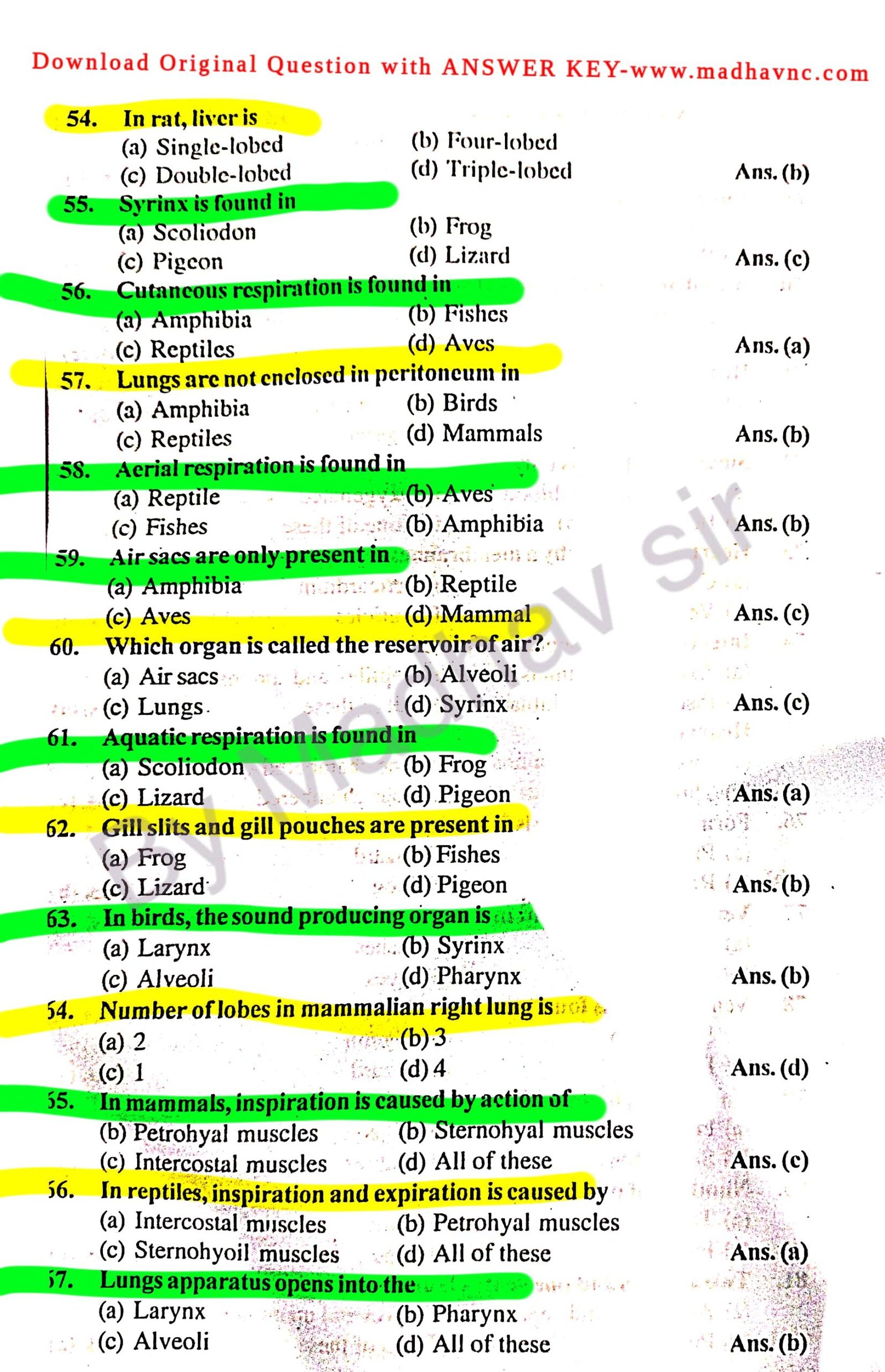
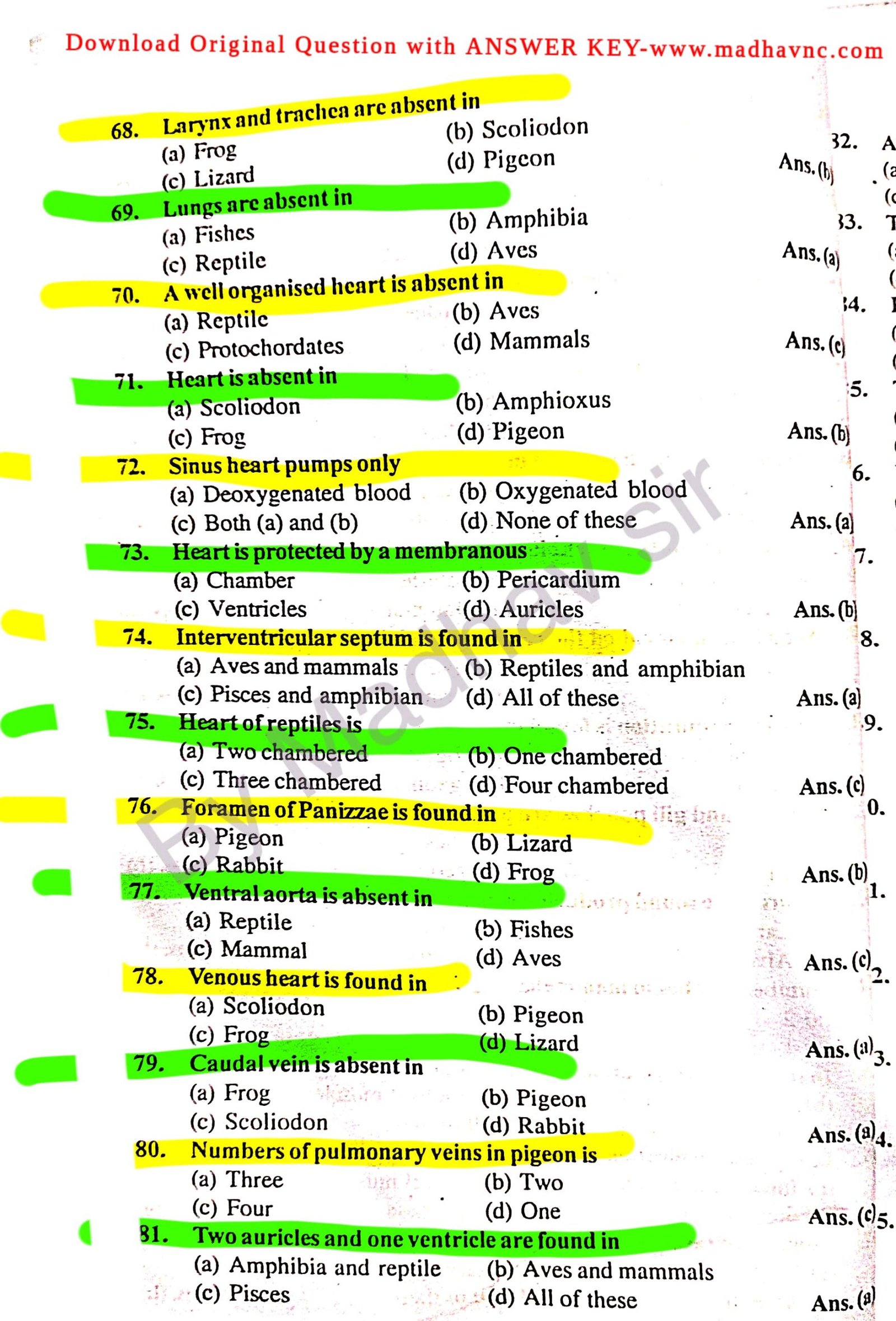
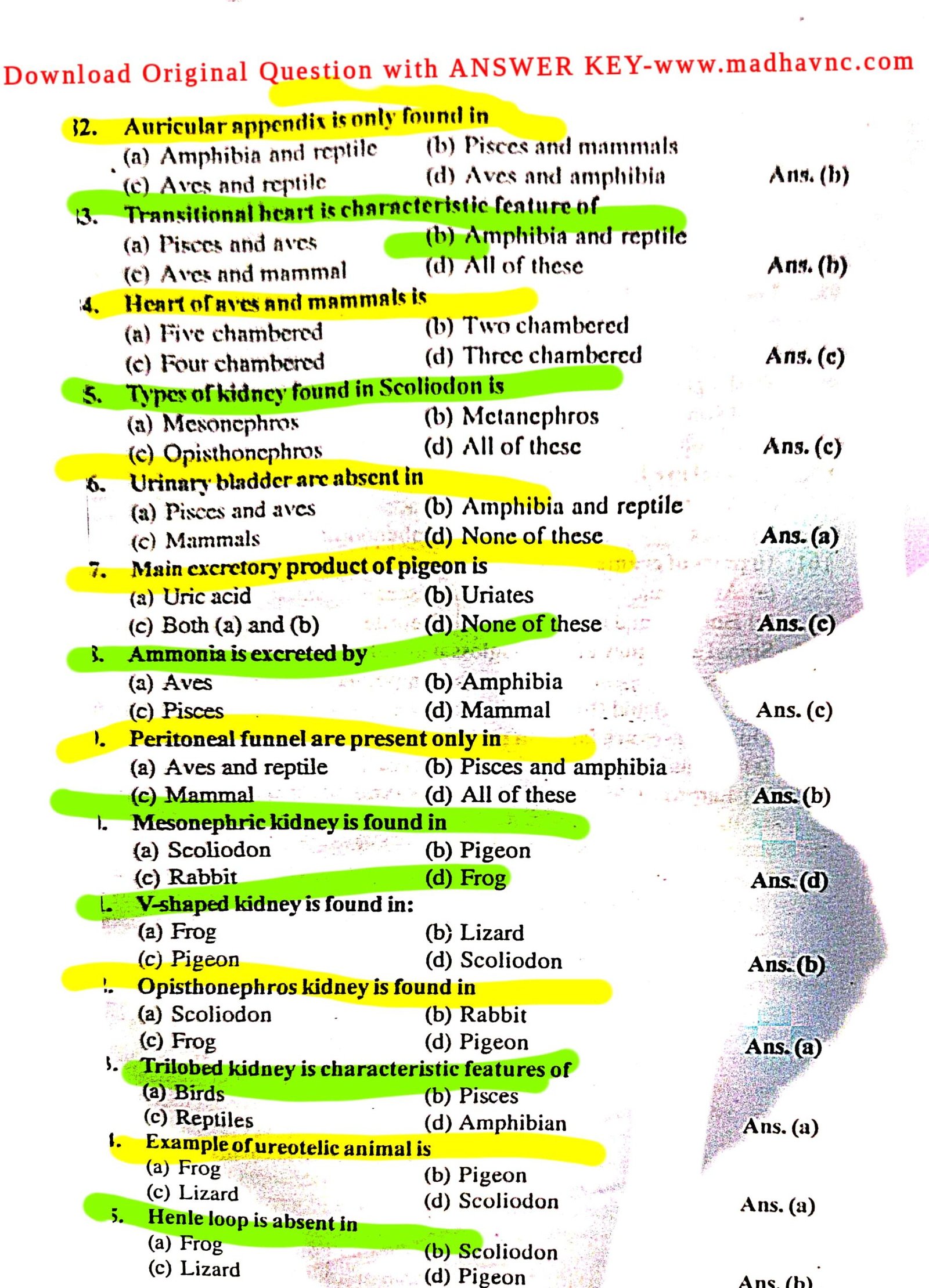
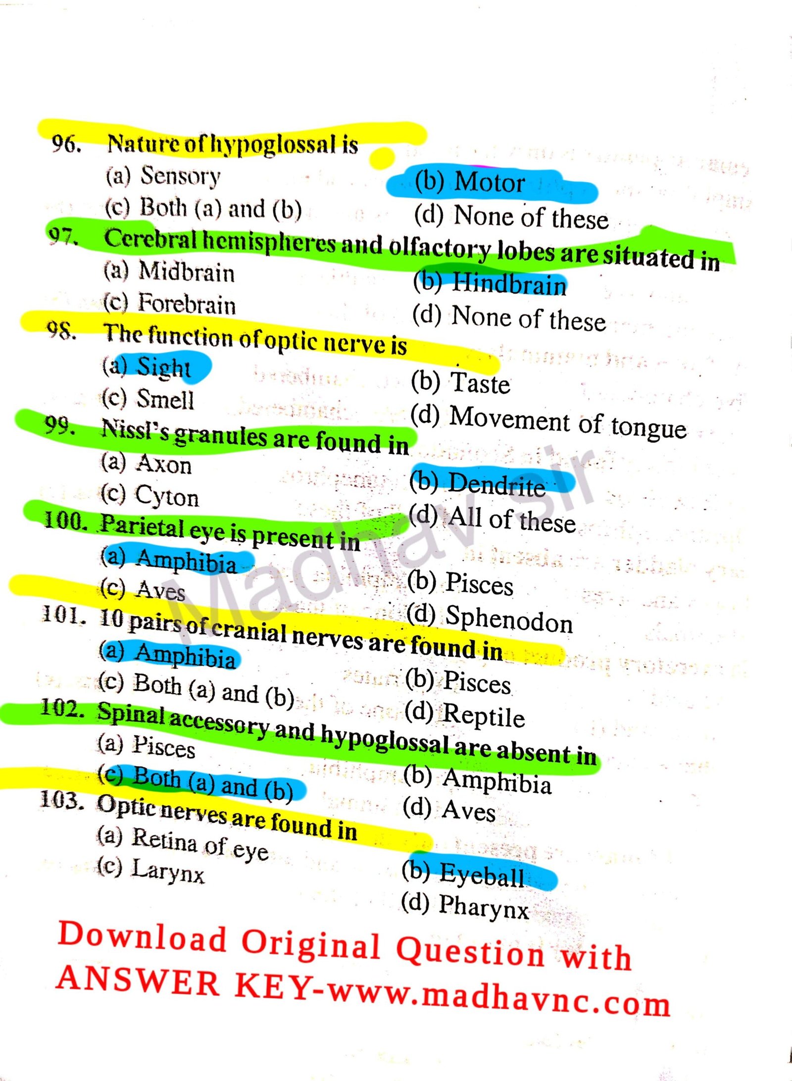
UG 3rd Sem Zoology MJC/MIC/MDC-3 Question Pepper 2025 | lnmu 3rd Sem Exam 2023-27 | Zoology MJC 3 Question Pepper

Join For Latest News
| Join Now | |
| Teligram | Join Fast |
| Join Now | |
| 3rd Sem Live Class | Join Channel |
lnmu 3rd Sem Exam 2023-27 | Zoology MJC 3 Question Pepper

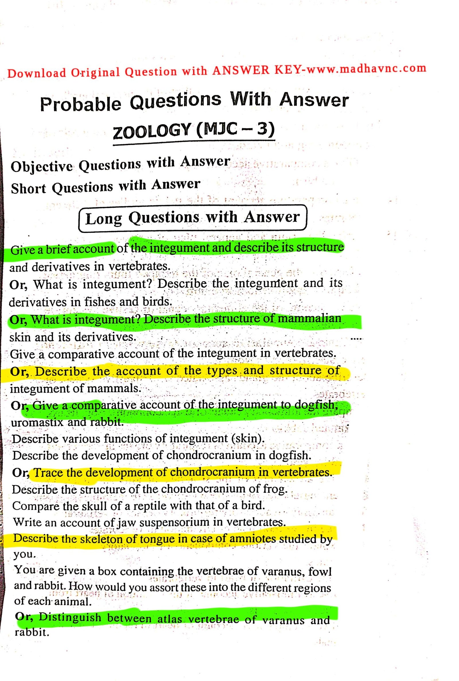
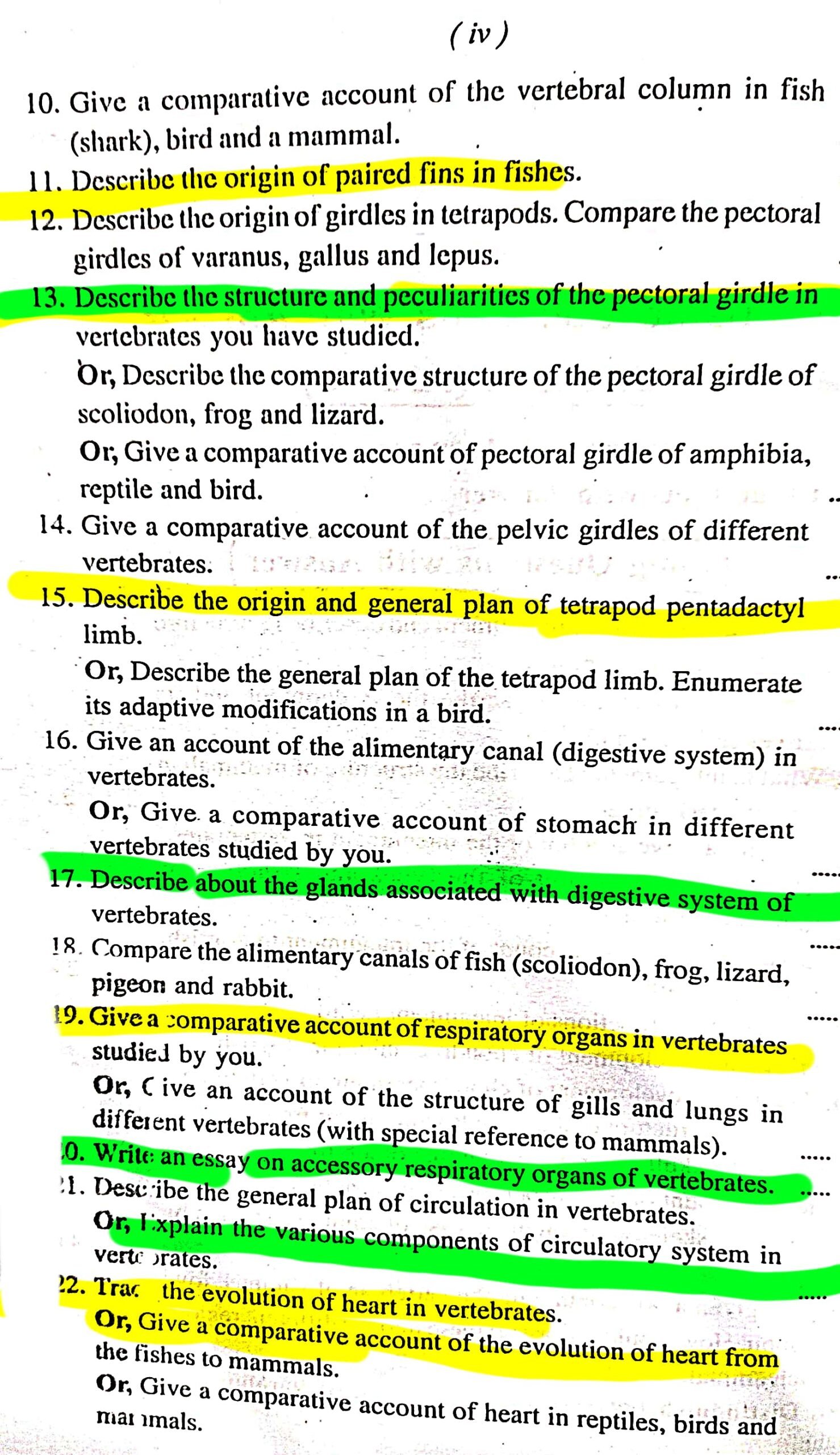
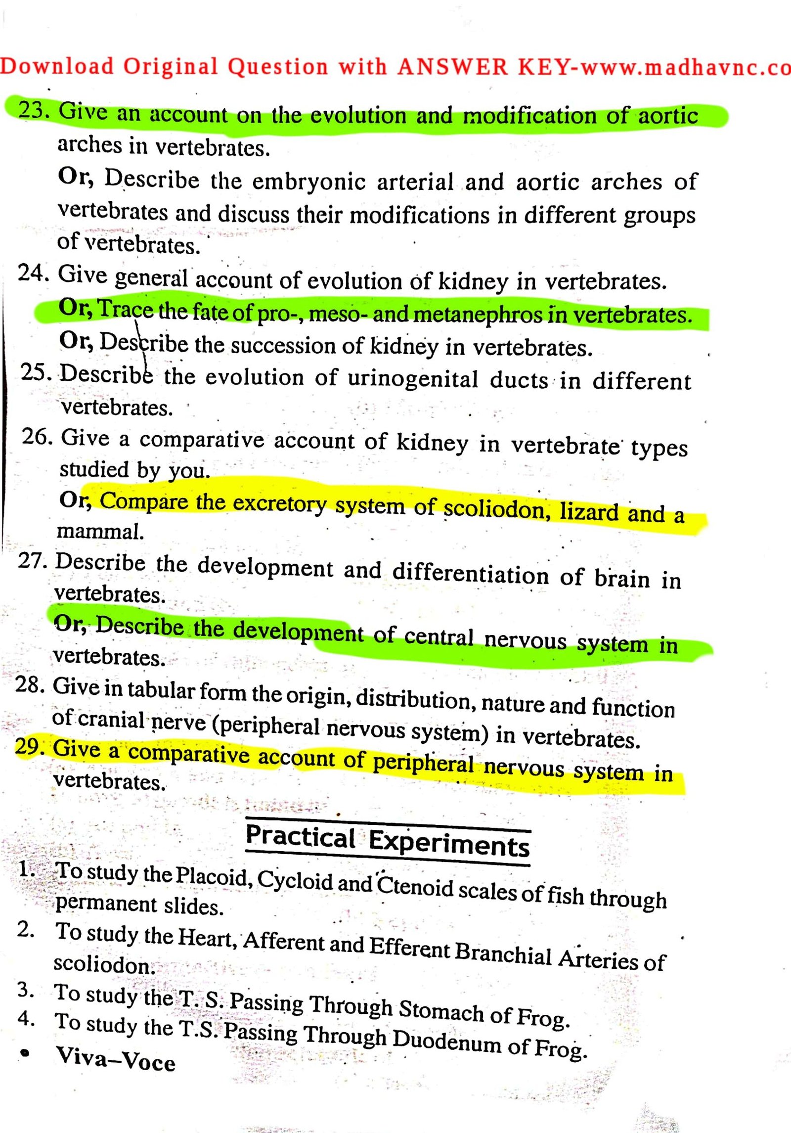
Confirm Subjective Questions Papper By Madhav sir
Q.1. Describe the general structure of integument.
Ans. The skin of all vertebrates is built according with the same basic lan. It is multicellular and differs from that of the invertebrates in having vo layers are as follows:
1. Epidermis: Epidermis is a stratified epithelium and normally quite in in comparison to dermis. It is further distinguished into two regions-) The outermost region of many layers of dead usually flattened (squamous) ells forms a horny, resistant covering or stratum corneum on the skin surface. s cells accumulate a horny protein, called keratin, gradually die and ventually wear off in the form of scurf or dandruff. Since keratin is tough and soluble in water, the keratinized stratum corneum provides protection gainst mechanical injuries, fungal and bacterial attacks and loss of body oisture. (ii) The innermost or basal region of epidermis includes a single w of living columnar cells, the Malpighian layer or stratum germinativum, nich is separated from the underlying dermis by a basement membrane. Its cells tively divide and continually replace the worn out cells of the cornified layer.
Epidermis is thin in aquatic vertebrates and rich in mucous glands. It is cker in land vertebrates and structures such as scales, feathers, hairs, nails, ws, horns and enamel of teeth are derived from its Malpighian layer.
2. Dermis: Dermis or corium, which is the inner layer of skin, is mparatively thicker than epidermis. It is composed of fibrous connective sue and contains many blood capillaries, lymph vessels, muscle fibres, rve fibres, sense organs and elastic fibres which bring the skin back to its rmal shape. Pigment cells or melanocytes are mostly located in dermis, hough sometimes pigment granules are also found in epidermis. Fat may cumulate as reserve food in special cells, called adipocytes, in deeper parts dermis and in the subcutaneous tissue. כת
Q.2. Give an account of the structure of mammalian hair.
Ans. Hair are complex cornified epidermal derivatives formed of ratinized epidermal cells, which are deeply sunken in the dermis and are closed in the tubular downgrowths of Malpighian layer. These are known hair follicles. The hair is distinguished into two parts: E
1. Hair Root: It is basal part of the hair, which is enclosed in the hair licle and lies embedded in the dermis. At the base of the follicle the hair ot is expanded into a bulb or inverted cup. It encloses the dermal papilla. e dermal papilla contains connective tissue, blood vessels and nerve fibres. e cells of hair root are living and possess power of division. These newly ided off cells add to the size of hair and gradually undergo keratinization.
Q.3. What is skeleton? Explain its various types.
Ans. The hardened tissues of the body together form the skeleton (scle = hard). Organism will remain small and slow moving if there had been skeleton for support and to serve as levers on which muscles can act. Skelet of invertebrates is most often secreted on the surface, forming a lifeless dead exoskeleton. Whereas skeleton of vertebrates develops most oft underneath the surface forming a living or growing endoskeleton.
The types of skeletons develop in vertebrates are as follows :
1. Epidermal horny exoskeleton: These include hard and horny
keratinized derivatives of epidermal layer of skin, such as claws, reptili scales, bird feathers and mammalian hairs, horns, nails and hoofs, etc. living amphibians lack an exoskeleton.
2. Dermal bony skeleton : Dermal bony skeleton is derived from
dermis of skin. It includes bony scales and plates or scutes (osteoderm finrays and antlers of fishes, reptiles and mammals. In fishes, dermal scal become exposed due to wearing out of epidermis and form exoskeleton.
Q:4. Describe various functions of endoskeleton.
Ans. Chief functions of endoskeleton can be enumerated as follows:
(1) To provide physical support to body by forming a firm and rigid internal framework.
(2) To give definite body shape and form..
(3) To protect by surrounding delicate internal organs like brain, heart, lungs, etc.
(4) To permit growth of huge body size (whale, elephant, extinct dinosaurs), since it is living and growing.
(5) To provide surface for attachment of muscles.
(6) To serve as levers on which muscles can act.
(7) To manufacture blood corpuscles in bone marrow.
****(8) To aid in hearing (ear ossicles).
(9) To help in breathing (tracheal rings, ribs).
Q. 5. Give an illustrated comparative account of the pectoral girdles of lasmobranch and bird.
Ans. Comparison of Pectoral Girdles of Elasmobranch and Bird
Elasmobranch (Scoliodon)
1.Pectoral girdle is cartilaginous.
2.Two halves of girdle join mid-ventrally but remain separate dorsally.
3.The dorsal end of each half arti-cùlates with the skull.
4.Each half is roughly semicircular piece of cartilage and is formed of two pieces:
Bird (Gallus)
1.Pectoral girdle is bony.
2.The two halves of the girdle are widely separated dorsally as well as ventrally.
3.
4.Each half is formed of a sabre-shaped scapula and a thick rod-like coracoid.
(i) Dorsal thick rod-like scapula and (ii) ventral thin and flattened coracoid.
5.At the junction of coracoid and scapula are three articular surfa-ces for the articulation of three basal cartilages of the pectoral fin.
5. There are no such articular surfaces. There is a glenoid cavity at the junction of two bones for the articulation of humerus bone of the fore-limb.
Q.8. Give a comparative account of oesophagus in vertebrates.
Ans. Oesophagus is a simple, muscular, distensible tube connecting pharynx with stomach. Its length is related to the length of neck. It is very short in neckless vertebrates (fishes and amphibians) but longer in amniotes, reaching extreme in birds, giraffes, etc. It may be lined internally with finger-like fleshy papillae (elasmobranchs), horny papillae (marine turtles) or longitudinal folds. In grain feeding birds (pigeon), oesophagus forms a paired or unpaired membranous sac or crop, modified for storage of food. In pigeons of both sexes, epithelial lining of crop undergoes fatty degeneration controlled by a pituitary hormone, prolactin, forming ‘pigeon’s milk’ which is fed to nestlings. Oesophagus has no serous coat as it lies outside coelom. In mammals, when it passes diaphragm it has serous lining. Food bolus passes down oesophagus into stomach by a muscular wave of contraction and relaxation called peristalsis. Oesophagus exhibit difference from rest part of the alimentary canal. The important differences are-it has no visceral peritoneum lining but outer covering is tunica adventitia. Muscle fibers of the anterior part of the oesophagus are striped, middle part is both striped and non striped and posterior part is only unstriped muscles. But ruminants all along their oesophagus have striped and voluntary muscles. Internal mucosal lining is of stratified squamous epithelial cells.
Q.9. Give a comparative account of pancreas in vertebrates.
Ans. Pancreas is also a constant structure of all vertebrates and second largest digestive gland after liver. Typically, pancreas arises as one or two ventral diverticula from liver bud and one dorsal diverticulum from embryonic duodenum. It is endodermal in origin from embryonic archenteron. Distal portions of diverticula divide to form acinous type glands, one dorsal pancreas and one ventral nancreas. Both may persist, as in fishes, but more generally two unite to form a single gland as in tetrapods. Proximal portions of Aerticula form pancreatic ducts all of which may persist. But usually the diet undergo reduction or fusion, so that only one or two pancreatic ducts rem in as in mammals. The ducts open into duodenum separately or jointly or quie of them may unite with the common bile duct.
Pancreas plays a dual role. It is partly exocrine secreting digestive enzymes through pancreatic ducts into duodenum and partly endocrine secreting hormones such as insulin. No pancreas is present in lancelet. In lampreys, some teleosts, lungfishes and lower tetrapods, it is distributed diffusely in liver, mesenteries and intestinal wall and probably only exocrine in function. In elasmobranchs and higher tetrapods, pancreas is well-defined and compact.
Q.11. Write a note on renal portal system…
Ans. The venous blood from each hindlimb is collected by a pair veins. The femoral collects blood from the muscles and skin on the outer sid of hindlimb and the sciatic from the muscles on the inner side. The femoral. on entering the body cavity, bifurcates into two branches. The outer branch. termed as renal portal vein and runs along the outer surface of the kidney its side. Here, it receives blood from the dorso-lateral body wall by dorse lumbar vein and finally ramifies into capillaries in the substance of kidne! The inner branch of femoral is known as pelvic vein. The pelvic veins either side unite mesially forming the anterior abdominal vein.
Q.12. Write a note on hepatic portal system.
Ans. The hepatic portal system includes veins which collect blood from e various regions of the alimentary canal. The oesophageal vein from the sophagus, gastric vein from the pancreas, splenic vein from the spleen and testinal vein from the small intestine collectively open into the hepatic rtal vein. It enters the left liver lobe and finally branches into capillaries.
A pair of hepatic veins emerge out of the liver and open into the post val, thus pouring the collected blood into it.
Significance of Hepatic Portal System: The digested food from the
mentary canal is absorbed into the blood and is carried to the liver where:
(1) Excess sugar is converted into glycogen for storage.
(2) Ammonia produced during the process of protein digestion is nverted into urea, which is carried to the kidneys.
(3) Fat particles are picked up by the Kupffier cells of liver.
(4) Undesirable substances such as carbolic acids, etc. are also extracted t by liver cells.
(5) Harmful solid particles like bacteria, etc. are removed and reacted on by the liver cells.
Confirm Subjective Questions Papper By Madhav sir
Q.1. Give a brief account of the integument and describe its structure nd derivatives in vertebrates.
Or, What is integument? Describe the integument and its derivatives in shes and birds.
Or, What is integument? Describe the structure of mammalian skin nd its derivatives.
Ans. Integument is the outermost covering of the body of vertebrates. It a multicellular and multilayered structure formed of an outer layer of idermis and an inner layer of dermis.
1. Epidermis: The epidermis is derived from the ectoderm of the embryo id is formed of several layers of stratified epithelial cells. The lowermost yer of the epidermis is formed of living columnar cells and is known as atum malpighi or stratum germinativum. Its cells possess power of division d proliferate towards its outer side. These move upwards and gradually atten. Their cytoplasm undergoes keratinization and becomes horny and > nucleus disintegrates. The outermost layer of these cells forms the stratum rneum. Its cells are horny, hard, flattened and dead.
2. Dermis: The dermis is comparatively thicker and is derived from the esoderm of the embryo. It is formed of an outer loose layer of stratum ongiosum and an inner dense layer of stratum compactum. It is formed of nite-collagen and yellow elastin fibres and smooth muscles embedded in > connective tissue mass. The matrix is traversed by blood capillaries, nerve res, lymph vessels, pigment cells, sensory cells and nerve endings.
A number of structures like hairs, feathers, scales, bony plates, glands, :., are also found embedded in the dermis. The pigment is contained in a ecial type of branched cells, the chromatophores. These form a continuous ver below the epidermis. The chromatophores are of following types:
(1) Melanophores having black or brown pigment,
(2) lipophores with yellow or red pigment,
(3) iridocytes with crystals of guanine.
Structures derived from the integument are of two types: Those derived om the epidermis and the others derived from the dermis.
[I] Epidermal Derivatives: The epidermal derivatives are of two types: